We use cookies to help us provide you with a more enhanced and personalized experience adapted to your interests. By using our site you agree to our Terms of Use and Privacy Policy, including our use of cookies.
A German manufacturer of stone-retrieval baskets works with a U.S. manufacturer of specialty optical fiber. The result is a basket that, thanks to its coaxially integrated optical fiber, can simplify and shorten minimally invasive urological surgery.
The treatment of kidney stones has changed dramatically over the years. Instead of open surgery, today minimally invasive endoscopic-based procedures can be used. Once a stone is found, it can usually be removed using a nitinol basket. If the stone is too far up the urinary tract, fragmentation using laser energy is used to pulverize it. Pulverization is achieved by the introduction of an optical fiber to deliver the laser energy. This procedure is called intracorporeal lithotripsy.
Pulverization using laser energy may vary. Combining a long pulse duration with low pulse energy and high pulse frequency will blast the stone into dust. The small dust particles are eliminated, naturally. But the high pulse energy will cause the ambient temperature to rise and may cause damage to surrounding tissue. An alternative to pulverization is fragmentation. Fragmentation uses laser energy with a short pulse duration, high pulse energy and low pulse frequency. The resulting fragments can then be captured using a stone-retrieval basket.
Usually the stone is fragmented prior to the pieces being captured by the basket. But sometimes, depending on the location of the stone, the reverse order is necessary. In these cases, where the stone is captured and then fragmented, there is the risk of the laser energy damaging the stone-retrieval basket as well as the surrounding tissue.
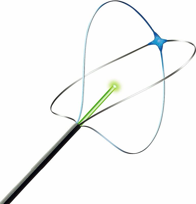
The next logical development in intracorporeal lithotripsy is an instrument that coaxially integrates optical fiber with the stone-retrieval basket. This improved instrument enables positioning of the basked and the optical fiber at the same time. The stone is safely trapped and fragmented without damaging surrounding tissue or the basket. Surgery time is shortened since only one instrument is needed.
This new device was developed by Endosmart GmbH in Stutensee, Germany together with OFS, a U.S. designer and manufacturer of specialty optical fiber.
A typical laser system for lithotripsy is based on Ho:YAG (Holmium:Yttrium-Aluminum-Garnet) laser which uses at a wavelength of 2123 nm with an average power of 30 W. Pulse duration, peak power and frequency are adjusted according to the individual treatment. For example, the laser pulse could be up to 18 kW peak power or 3.5 J pulse energy. To enable orientation of the instrument, the system delivers a visible red or green pilot light.
Light is guided even under extreme bending
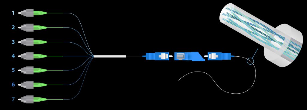
The step-index multimode optical fiber used to guide the laser can have a pure silica core and a fluorine-doped glass cladding or a Germanium-doped core with a pure silica cladding. The different refractive indices of core and cladding enable the laser to propagate longitudinally in the fiber core. For guiding the light under extreme bending, an additional UV cured fluoroacrylate coating is applied. The fluoroacrylate coating has a lower refractive index than either of the glass claddings and acts as a secondary cladding for guiding the light. The optical fiber that is used with the nitinol basket described above has a core diameter of 272 µm and a silica cladding diameter of 299 µm. Around that, a 330 µm UV cured fluoropolymer coating is applied acting as a second optical cladding and finally, an ETFE buffer of 400 µm is applied.
Glass fibers are also used for medical diagnostics. Current developments are focused on simultaneous diagnosis and treatment.
Researchers at the ARC Centre for Excellence in Nanoscale BioPhotonics (CNBP) discovered an exciting new method that could make it possible to use 3D microscopy to view hard-to-reach areas of the human body. This method uses fiber optic bundles to miniaturize a type of 3D imaging called “light field imaging.” Taken to extreme new levels, this imaging could make in-body use possible.
This method could find widespread use in diagnostic procedures called optical biopsies. In these biopsies, suspicious body tissue is investigated using medical endoscopic procedures.
Until now, light field imaging could be performed only with bulky hardware such as camera arrays or modified consumer cameras. Instead of trying to shrink existing equipment, the researchers realized that fiber optic bundles already used routinely in microendoscopy were actually suitable light field imaging devices.
Fiber optic bundles are groups of tens of thousands of microscopic optical fibers. Each fiber in the bundle acts like a pixel in a camera. The result produced is a 2D image that is transmitted through the fiber bundle.
Along with recording a 2D picture, light field imaging systems also measure the incoming angles of all light rays in the picture. With this information, doctors can map the picture in stereo 3D in the same way that humans perceive depth. According to the researchers, the primary challenge will be how to record this angular light ray dimension that is often hard to capture.
According to Dr. Antony Orth, project lead and Research Fellow at the RMIT University node of the CNBP, “The key observation we made is that light ray orientation information is actually transmitted by the fiber optic bundles to the microendoscope. You just need to know what to look for and how to decode it.”
Dr. Orth believes that, given the right mathematical framework, researchers can decode the patterns, transform them into a light field, and do incredible things such as refocusing, depth mapping and visualizing the image in stereo 3D. He believes that this light field technology could potentially bring an entirely new depth dimension into optical biopsies. This capability would let doctors examine suspect tissue without removing a sample from the patient.
The research group is meeting with physicians to discuss how to test this technique in medical clinics and to also identify the medical procedures most likely to benefit from 3D visualization in the microscale.
A new, air-filled optical fiber bundle could dramatically improve medical endoscopes. This technology could also help create endoscopes that produce images using infrared wavelengths. If so, this breakthrough would allow diagnostic procedures that aren’t currently possible.
In the Optical Society (OSA) journal Optics Letters, University of Bath (U.K.) researchers showed that these new fiber optic bundles (called air-clad imaging fibers) deliver resolution equal to the best commercial imaging fibers. And the bundles do this at twice the wavelength range of these fibers. Because of this, these air-clad imaging fibers could help create new, smaller endoscopes with even better resolutions.
HOW ENDOSCOPES WORK
Used in minor surgery and imaging, endoscopes use bundles of optical fibers to obtain images from inside the body. Light that falls on one end of the fiber bundle travels through each fiber to the far end. This process sends a picture as thousands of spots, much like pixels in a digital picture.
TESTING THE BUNDLES
Instead of using cores and claddings of two types of glass, the new bundles use an array of glass cores covered by hollow glass capillaries filled with air. These air-filled capillaries act as the fiber cladding.
To test the imaging fibers, the research team created an air-clad fiber bundle. This bundle matched the resolution of a leading commercial fiber (with the same spacing between cores). The team then stacked multiple, smaller honeycomb structures to place more than 11,000 cores into the fiber.
The researchers used the air-clad fiber bundle and the commercial fiber to image a standard test target image. And the result? The air-clad fiber performed well beyond the wavelength range that a visible camera could detect. And when the researchers switched to an infrared camera, the fiber created a clear image at twice the wavelength that the commercial fiber reached.
REAL-WORLD USE OF FIBER BUNDLES
Along with medical diagnosis and care, the new optical fiber bundles could be used for industrial applications. These uses include monitoring the contents of hazardous machines and viewing the inside of oil and mineral drills. These types of fibers are becoming more and more popular for a variety of purposes.
OFS Laboratories, one of the world’s leading optical fiber research labs, and the research arm of OFS, has performed major work in this area. The development of Microstructure Optical Fibers (MOFs) is one result of this work. The MOFs created by OFS Labs are a new class of optical fibers that are substantially different from conventional optical fibers.
Placing sensors inside the human body can help researchers and physicians to understand and treat a variety of medical conditions. However, while implanting a sensing device may be routine, having it remain in the body long enough to perform its job and then be safely removed is an entirely different and significant challenge.
Now a team of Italian and Greek researchers have embedded fiber Bragg gratings, a type of device that reflects certain light wavelengths and can be used as a sensor, inside of dissolvable optical fibers. This new technology may allow the long-term monitoring of the biomechanical and chemical properties of various organs and anatomical features inside the body.
Fiber Bragg gratings placed into optical fibers are routinely used to measure stresses placed on bridges, commercial airliner wings and other areas where detailed, real-time monitoring is critical. The newly-developed fiber Bragg gratings are able to break down, similar to absorbable sutures and, because they have been embedded into optical fibers that are also bioresorbable, they should be safe for use inside the body. Ideally, they would be implanted, left inside the body to perform sensing and eventually disappear completely without the need for removal. (more…)
A new fiber optic technique for assessing muscle health could eliminate the need for painful muscle biopsies. To diagnose a muscular disorder, disease or infection, physicians must often extract a tissue sample. However, these biopsies can be quite painful and difficult to perform.
A story in Medical XPress reports that researchers at the Rehabilitation Institute of Chicago (RIC) have developed a less invasive alternative that uses a thin fiber optic probe to quickly scan and measure the health of muscle tissue. And, for the first time, the team has now tested the system on living muscle.
To read the article, please go here. To access the full report in Biophysical Medical, use this link. For information on OFS fiber solutions for medical devices, please click here.
Medical imaging faces limitations inherent to its mode of presentation. While computer models and virtual reality are much more effective than 2D depictions, the result continues to be still images on a computer screen. Even with stereoscopic techniques, a user’s ability to visualize the result can depend on using a keyboard or mouse to interpret the model. And, with 4D experimental medical data (such as MRI), objects are displayed as computer animations or static pictures.
A recent Biophotonics article by Thomas Britton and OFS’ Jaehan Kim shows how a hands-on, 3D-printed brain model equipped with optical fibers can help clinicians and patients to visualize brain function activity while avoiding the shortcomings of 4D neuroimaging techniques.
OFS will showcase its new Shape Sensor Fiber at the BIOS/Photonics West Exposition in San Francisco, January 28-February 2, 2017.
To create the Shape Sensor Fiber, OFS developed a technology platform to produce high-quality, twisted multicore optical fiber with continuous Fiber Bragg Gratings (FBGs). This type of fiber with FBGs provides stable and good signal-to-noise ratio throughout the fiber length and ease of use to customers. The manufacturing platform also allows OFS to customize and optimize the fiber to meet various customer demands more economically. In addition, OFS also offers low back reflection distal termination, multicore connectorization and fan-outs to support customer demand.
Many medical device companies are developing cutting-edge endoscopes, catheters and other equipment with shape sensing technology to increase the quality of patient care. By embedding or surface-attaching the fiber to surgical tools or other devices, technicians can calculate and reconstruct the 3D shape of an instrument on a display screen. By allowing users to monitor the exact shape and position of the instrument, physicians can conduct minimally invasive surgery (MIS) or treatment which generally results in shorter recovery times, less pain and trauma, reduced rates of infection and shorter hospital stays.
In a recent study, researchers from the University Hospital Jean Minjoz (Besacon, France) demonstrated that optical coherence tomography (OCT) imaging can more readily visualize the coronary arteries in patients undergoing percutaneous coronary intervention (PCI) and lead to better outcomes when compared to standard angiography-guided PCI.
The study found that OCT provided useful additional information beyond that obtained solely by angiography, and impacted directly on physician decision-making. In fact, the use of OCT led to a change in procedural strategy in half of the cases.
In cardiology, the use of OCT involves introducing a miniature fiber optic catheter into the coronary artery to check vessel size, lesion traits and both stent positioning and expansion. OCT is also used in ophthalmology to assess the progression of macular degeneration, glaucoma and other ocular diseases.
To access details of the study, please go here and also here.
The physical characteristics of high-quality, silica optical fiber make it a natural choice for a broad range of uses, including many in the medical industry. For example, fiber can provide a very compact, flexible conduit for light or data delivery in equipment, surgical and instrumentation applications.
However, users must carefully choose the right optical fiber to avoid delays in product design and launch, along with increased development costs. A recent Medical Design Briefs article by OFS’ Jaehan Kim and Jonathan Loft explores the wide array of fibers available for this market. To access this article, please go HERE.
The commercial use of optical fiber in harsh environments is continually growing. These applications include medical probes that undergo sterilization at elevated temperatures and distributed sensors in oil and gas pipelines and wells exposed to extreme heat and cold. For these fibers to be used successfully, researchers and manufacturers must address the issues of fiber performance and reliability under the harshest conditions.
However, current theories and knowledge on the strength and dependability of silica-based optical fiber have been based almost exclusively on experiments conducted in optical telecommunications environments. Moreover, these tests only used a relatively narrow range of temperatures. For usage in extreme environments, fiber developers and users need new data and information.
In a recent white paper from OFS Specialty Photonics, researchers describe a setup for testing the tensile strength of optical fiber when exposed to high temperatures. This paper also reports the initial results of dynamic tensile strength testing conducted on polyimide-coated optical fiber at elevated temperatures over various time intervals.
To learn more and access this white paper, CLICK HERE.

 Researchers at the ARC Centre for Excellence in Nanoscale BioPhotonics (CNBP) discovered an exciting new method that could make it possible to use 3D microscopy to view hard-to-reach areas of the human body. This method uses
  A new, air-filled optical fiber bundle could dramatically improve medical endoscopes. This technology could also help create endoscopes that produce images using infrared wavelengths. If so, this breakthrough would allow diagnostic procedures that aren’t currently possible.
 Placing sensors inside the human body can help researchers and physicians to understand and treat a variety of medical conditions. However, while implanting a sensing device may be routine, having it remain in the body long enough to perform its job and then be safely removed is an entirely different and significant challenge.
 A new fiber optic technique for assessing muscle health could eliminate the need for painful muscle biopsies. To diagnose a muscular disorder, disease or infection, physicians must often extract a tissue sample. However, these biopsies can be quite painful and difficult to perform.